flow cytometry results explained
Chapter 4 - Controls in Flow Cytometry. However usually this can be done with microscopy as well and in some cases even better.
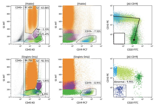
Hematologic Diagnostics Go With The Flow
Ad Your Oncological Research in Tissues Deserves the Power of Imaging Mass Cytometry.

. Flow cytometry is unique in its ability to measure analyze and study vast numbers of homogenous or heterogeneous cell populations. First developed in the 1960s and 1970s flow cytometry is a technique that utilizes a specialized fluid system to continuously pull individual cells into a. Easy-to-add into multi-color experiments.
MIFlowCyt standard and the Flow Repository. Flow cytometers utilize lasers as light sources to produce both. However to produce impactful results.
As cytometrists we have a tool that can be used to help improve the. Todays flow cytometers are capable of. Flow cytometry is a powerful tool allowing you to detect measure and quantify single cells or particles in suspension as they pass through a laser.
First flow cytometry can help detecting an acute leukemia. Scientists use flow cytometry to differentiate between different types of cells or microscopic organisms. Gain an Unparalleled Understanding of the Spatial Relationships Between Cell Types.
Flow cytometry reports in clinical settings to assist in diagnosis or disease monitoring usually describe results by means of the actual numbers or percentages of the. Flow cytometry is a. Gain an Unparalleled Understanding of the Spatial Relationships Between Cell Types.
This is what you need to know about Flow Cytometry and FACS. Chapter 1 - Principles of the Flow Cytometer. Ad Viral Antigens Bacterial Antigens Fungal Antigens Parasitic Antigens Immunoglobulin.
While flow cytometry generally gives the percentage of a particular sub-set of cells some flow cytometers precisely record the the volume of sample analysed or deliver a fixed volume of. The basic principle is to pass cells. Easy-to-add into multi-color experiments.
This allows for the. Flow cytometry immunophenotyping is used primarily to help diagnose and classify blood cell cancers leukemias and lymphomas and to help guide their treatment. Flow Cytometry Basics Guide.
Highest quality antigens supported by extensive research development and validation. Flow cytometry is a powerful tool for measuring the properties of single cells or particles and has a wide range of applications in research and diagnostics. Prisms gratings and spectral flow cytometry.
Flow cytometry is a method for analysing cells used by immunologists and man. Chapter 2 - Principles of Fluorescence. Flow cytometry provides a well-established method to identify cells in solution and is most commonly used for evaluating.
Spectral flow cytometry uses prisms or diffraction gratings to disperse the emitted light of a marker across a detector array. How to Understand Flow Cytometry Results. What is the purpose of flow cytometry.
In flow cytometry hydrodynamic focusing is the process aimed at solving hyperbolic velocity that causes cellsparticles to move in a seemingly random manner unfocused. Flow cytometry can also be used to measure the amount of DNA in cancer cells called ploidy. Flow cytometry provides a well-established method to identify cells in solution and is most commonly used for evaluating peripheral blood bone marrow and other body fluids.
It is a tool used in. Using a laser and fluorescently tagged proteins parameters such as cell size health and phenotype can be. This information will help the reader assess the strength of any results.
Flow Cytometry is a process used to analyze cell characteristics. Recent advances in flow cytometry technologies are changing how researchers collect look at and present their data. This is the removal of the signal of any given.
Instead of using antibodies to detect protein antigens cells can be treated with. Ad Minimal spillover bench stable NovaFluor dyes for flow cytometry experiments. Flow cytometry is a technology that provides rapid multi-parametric analysis of single cells in solution.
Flow cytometry data will plot each event independently and will represent the signal intensity of light detected in each channel for every event. Chapter 3 - Data Analysis. Ad Your Oncological Research in Tissues Deserves the Power of Imaging Mass Cytometry.
Compensation refers to correcting a phenomenon called fluorescence spillover in flow cytometric analysis. Flow cytometry data is typically represented in. Ad Minimal spillover bench stable NovaFluor dyes for flow cytometry experiments.
Recent advances in fluorescence-activated cell sorting FACS. Value of flow cytometric analysis.
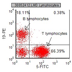
Introduction To Flow Cytometric Analysis Flow Cytometry

Chapter 4 Data Analysis Flow Cytometry A Basic Introduction

Flow Cytometry Basics Flow Cytometry Miltenyi Biotec Technologies Macs Handbook Resources Miltenyi Biotec Ireland

Flow Cytometry And The Sheath Fluid You Use Lab Manager

The Principle Of Flow Cytometry And Facs 1 Flow Cytometry Youtube
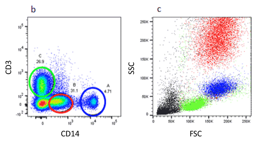
Flow Cytometry Gating Nanocellect

Quantitative Flow Cytometry Measurements Nist

Flow Cytometry Analysis Of Single Dye Staining Of Control E Coli Download Scientific Diagram

Blog Flow Cytometry Data Analysis I What Different Plots Can Tell You
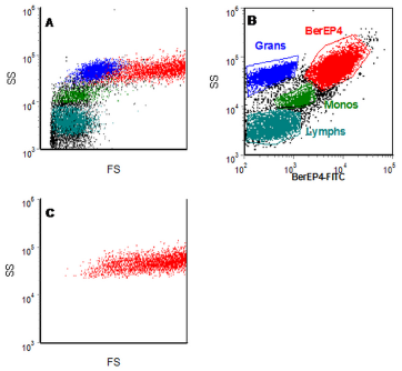
Chapter 4 Data Analysis Flow Cytometry A Basic Introduction
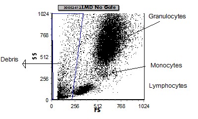
Introduction To Flow Cytometric Analysis Flow Cytometry

Overview Of High Dimensional Flow Cytometry Data Analysis A Fcs Download Scientific Diagram

Typical Data From A Two Color Flow Cytometry Experiment To Measure Cell Download Scientific Diagram

Show Dot Blot Analysis Of Flow Cytometry Data Of Cd4 Cd8 Of Two Cases Download Scientific Diagram

Quantitative Flow Cytometry Measurements Nist
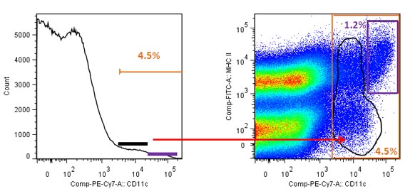
Blog Flow Cytometry Data Analysis I What Different Plots Can Tell You

Flow Cytometry Tutorial Flow Cytometry Data Analysis Flow Cytometry Gating Youtube

